
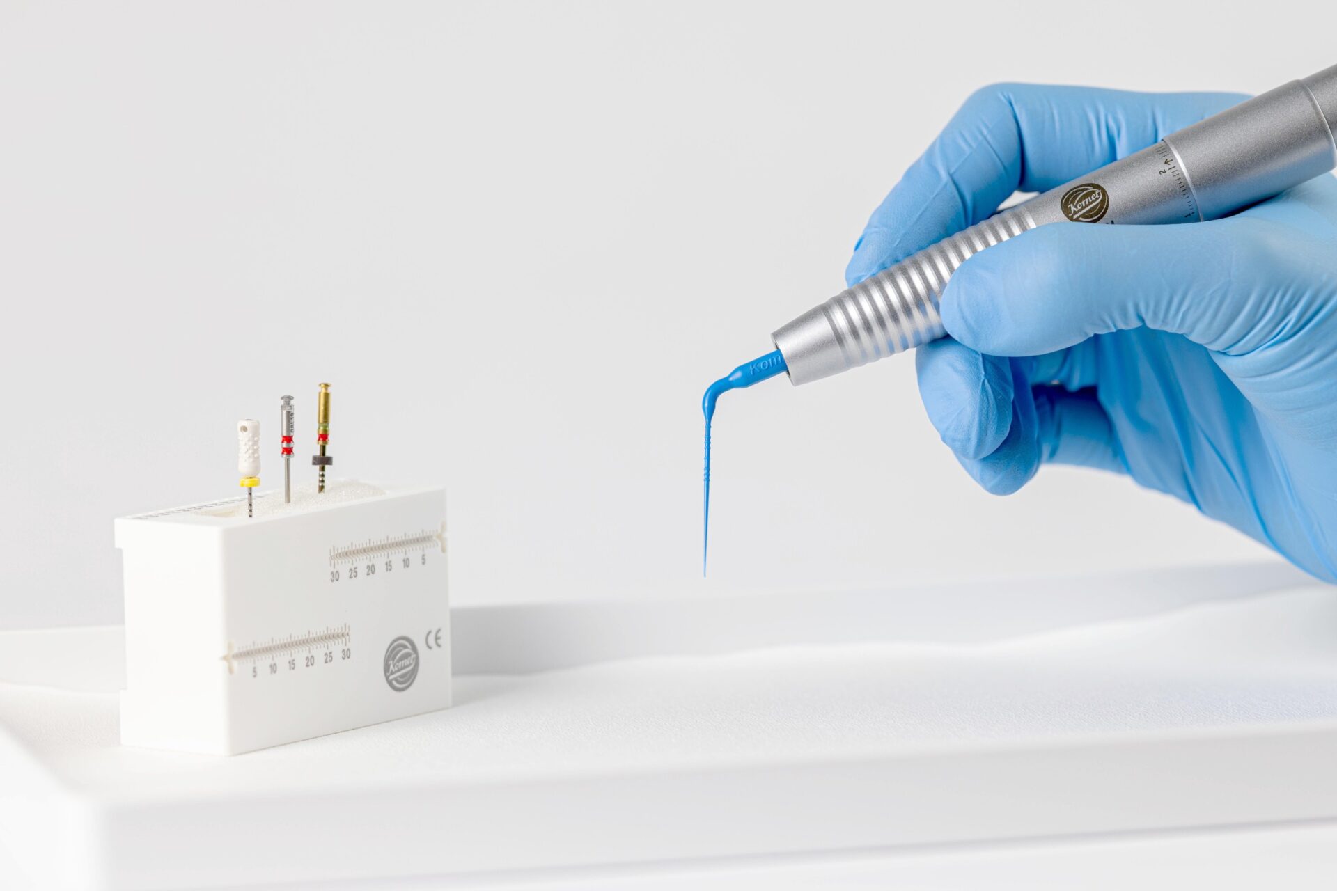
Komet BlueTip: High contrast, powerful cleaning performance.
Despite meticulous preparation of the access cavity and thorough treatment of all root canal sections using modern NiTi systems, significant portions of the root canal surface remain untreated, depending on the canal geometry. Therefore, the success of endodontic treatments depends largely on chemical disinfection. This is particularly true in cases of severely infected root canal systems.
Various methods are available to enhance the effectiveness of established rinsing solutions. Sonic irrigation has proven effective when it comes to performing high-quality endodontic treatments in everyday practice in an efficient, simple and cost-effective manner. The following case studies demonstrate the clinical benefits of the BlueTip (Komet) for a range of indications.
Case 1 – Primary treatment of
tooth 23
A 32-year old female patient came to my office with spontaneous, heat-induced and persistent pain in tooth 23. Clinical and X-ray examinations revealed a distal cavity (Fig. 1) without pathological probing depths. The cold test provoked a highly painful reaction that persisted beyond the stimulus. Irreversible pulpitis was diagnosed and, following appropriate patient education, the decision was made to perform a vital extirpation.
After administering a local anesthetic and isolating the area with a rubber dam, the caries was completely removed, exposing the inflamed pulp. To ensure a low-germ working environment and to monitor the rinsing liquids, a pre-endodontic build-up with composite was carried out prior to instrumental treatment. A manual glide path was then created and a needle-contrast image was taken.
Mechanical preparation took place using the FQ system (Komet) up to the master file 35/.06. After each sequential file application, the canal was irrigated with 6 % NaOCl and the rinsing solution was activated with the BlueTip (Figs. 2 and 3). Final disinfection was carried out using 17 % EDTA and three 30-second applications of 6 % NaOCl under ultrasonic agitation. The obturation was performed with BioSeal (Komet) and gutta-percha, employing the warm vertical condensation technique (Fig. 4). Finally, the coronal seal was created with composite.
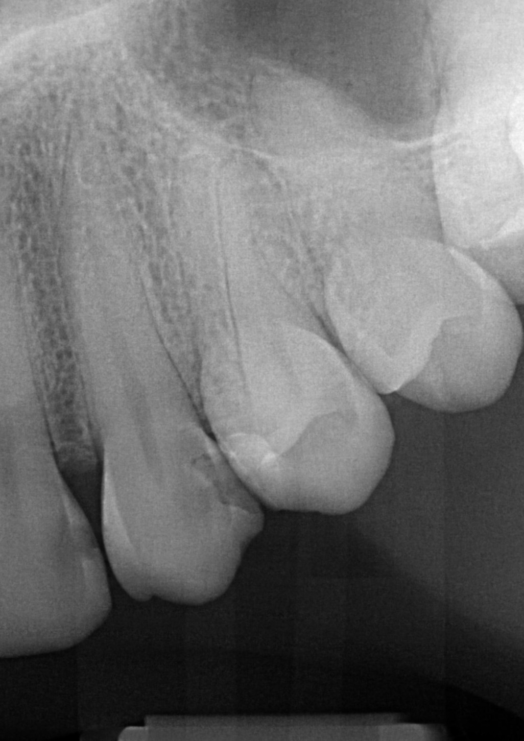
Fig. 1: Preoperative radiograph of tooth 23 showing distal coronal radiolucency
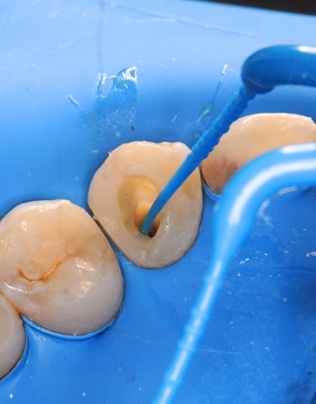
Fig. 2: Intraoperative use of the BlueTip (Komet) during the preparation of the root canal – palatal view
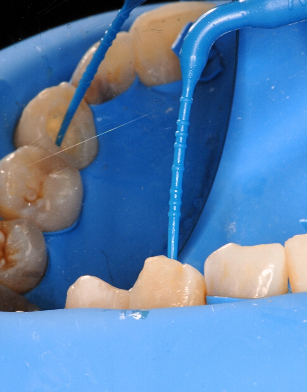
Fig. 3: Intraoperative use of the BlueTip (Komet) during the preparation of the root canal – labial view
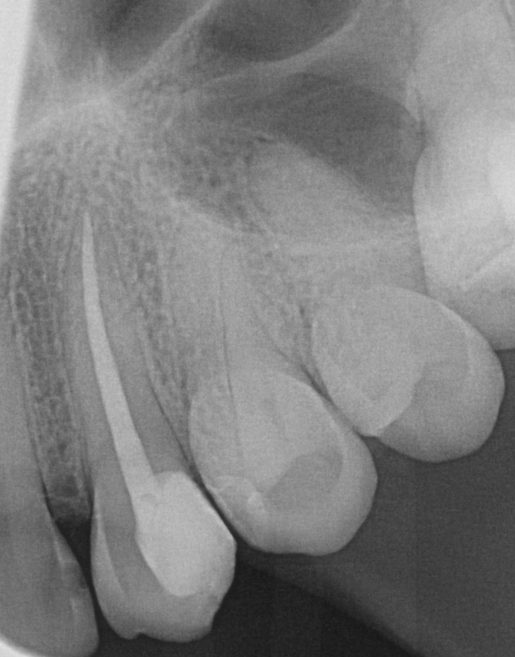
Fig. 4: Postoperative radiograph of tooth 23
Case 2 – Orthograde retreatment of tooth 16
A 27-year-old female patient came to my office two years after undergoing primary root canal treatment on tooth 16, with increasing discomfort. Radiographs showed periapical radiolucency at the palatal root, a misaligned root filling, and an untreated second mesiobuccal canal (Fig. 5). Following diagnosis and patient education, an orthograde retreatment was performed.
After administering a local anesthetic and isolating the area with a rubber dam, the access cavity was prepared and the filling material from the initial treatment was completely removed from the root canal with NiTi instruments. This was followed by the creation of a glide path in the second mesiobuccal canal. The canals were completely prepared with the FQ system.
The area was regularly rinsed with 6 % NaOCl throughout the entire preparation, and the rinsing solution was activated using the BlueTip. Once the shaping process was completed, the smear layer was removed with 17 % EDT. This was followed by thorough rinsing with NaOCl, during which the rinsing liquid was sound agitated several times. The root canal was obturated using BioSeal and gutta-percha employing the warm vertical technique.
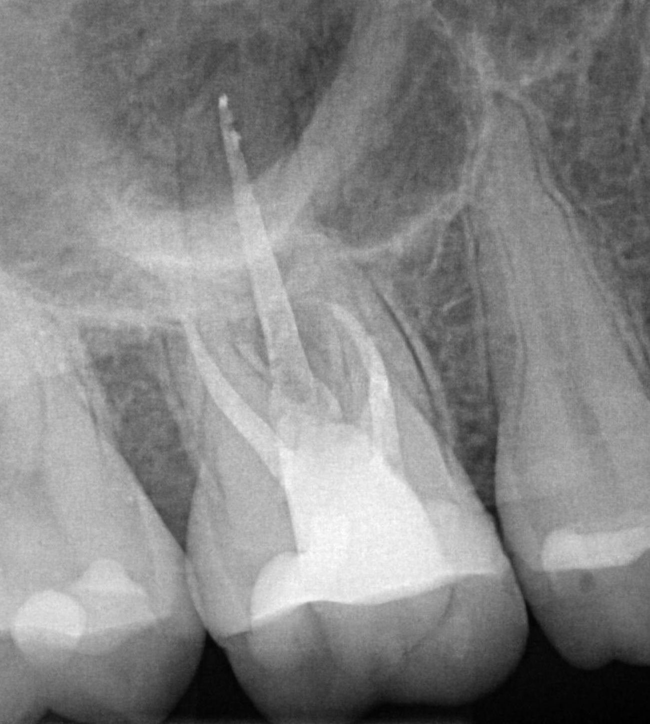
Fig. 5: Preoperative radiograph of tooth 16 showing periapical radiolucency at the palatal and mesiobuccal roots
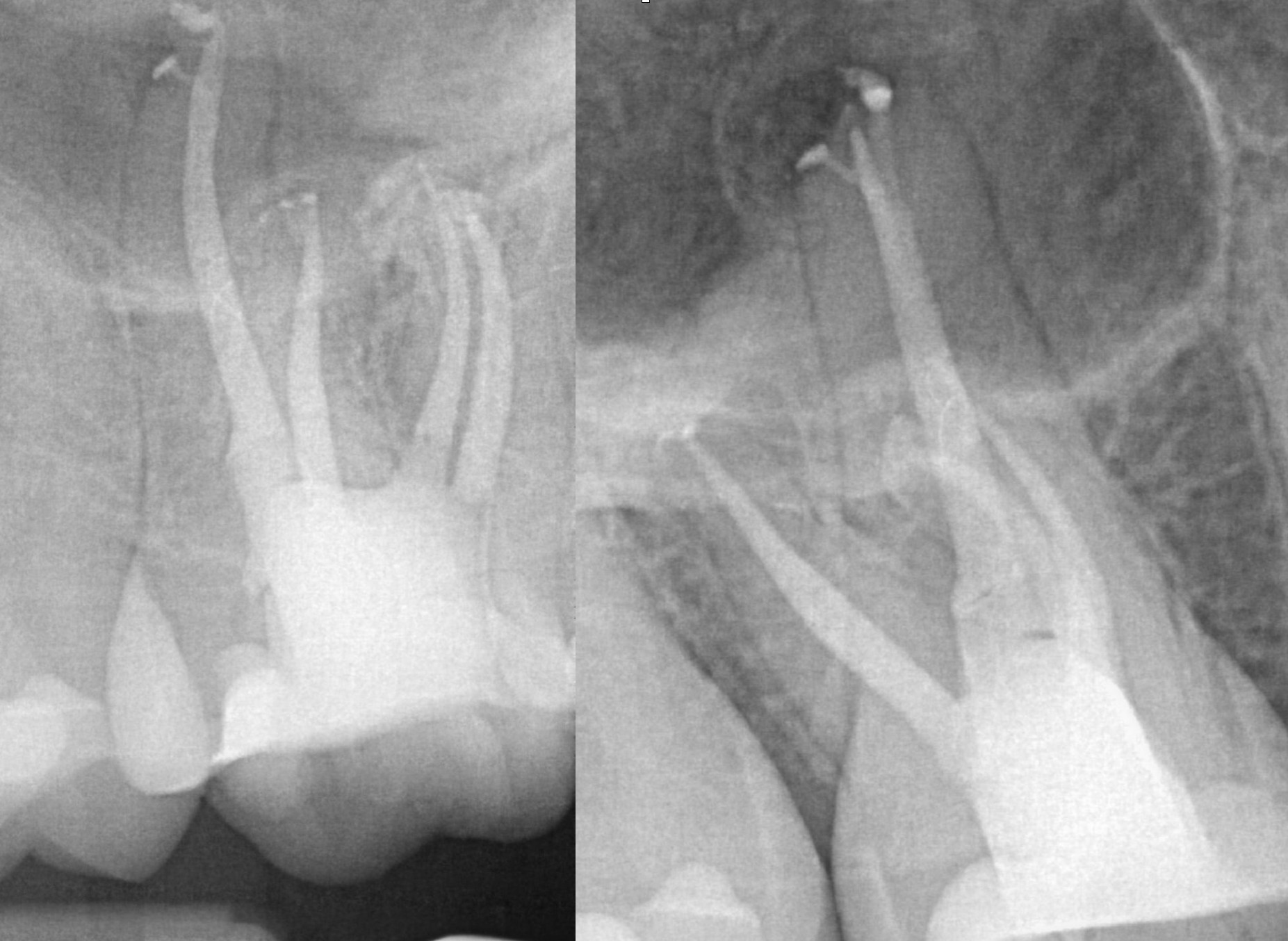
Fig. 6: Distal (left) and mesial (right) eccentric postoperative radiograph
The distal and mesial eccentric final radiograph (Fig. 6) showed precise obturation of the apical delta in all four canals. This indicates deep penetration of the rinsing liquids into the root dentin, even in the apical regions – an effect that can only be achieved through advanced agitation methods. The clearly visible length markings on the BlueTip significantly facilitated precise clinical use. The patient was pain-free after the first session.
Case 3 – Primary treatment of
tooth 17 involving the removal of a fragment
A patient came into my office having undergone a root canal treatment of tooth 17 that had been initiated elsewhere. The patient was referred to me because a mechanical glide path file had fractured in the apical third of the first mesiobuccal canal (Fig. 7). Although it was possible to pass the fragment, it could not be removed initially.
Once all canals had been fully prepared – including the second mesiobuccal canal which appeared to merge with the first canal in the apical third – the final disinfection took place as described in the previous cases. Thanks to the sonic activation of the rinsing solution in the second mesiobuccal canal with the BlueTip, the fragment in the first mesiobuccal canal could be dislodged and transported towards the crown, where it was successfully removed (Fig. 8).
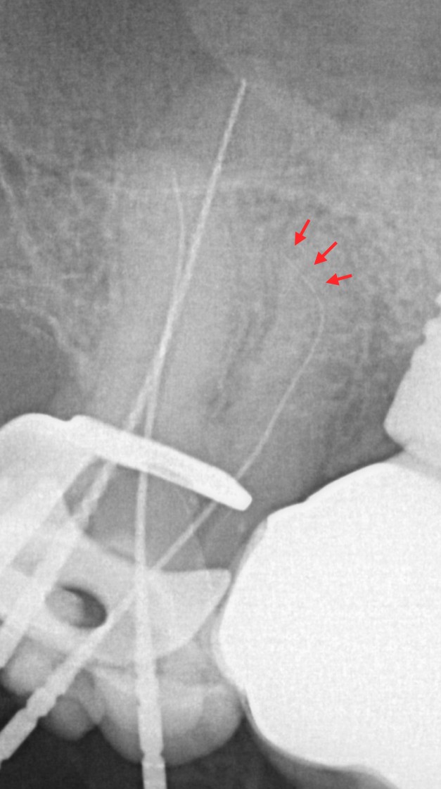
Fig. 7: Needle contrast image of tooth 17 showing the highlighted fragment in the first mesiobuccal canal. The discontinuity between the fragment and the inserted file is clearly visible.
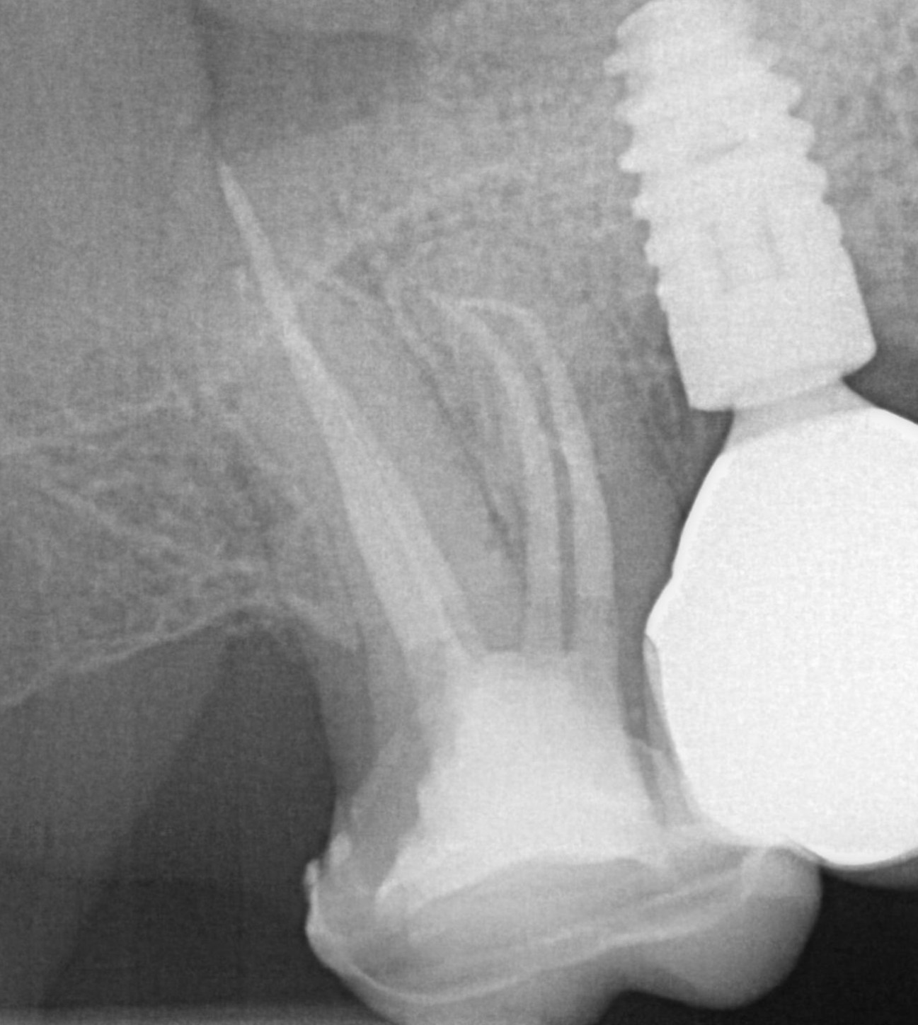
Fig. 8: Postoperative radiograph of tooth 17 after removal of the fragment and confluence between the first and second mesiobuccal canal
Conclusion
The agitation of rinsing liquids constitutes an indispensable part of modern endodontics. It facilitates preparation, reduces preparation errors and improves the penetration of rinsing solutions into the root dentin, resulting in significantly improved germ reduction. In some cases, it can even resolve instrument-related complications. The BlueTip is an indispensable tool for high-quality root canal treatments that can be effectively integrated into everyday practice.
About the dentist
Dr. med. dent. Laurens Intert
From the very beginning of his dental studies, which Dr. med. dent. Laurens Intert began in 2013 at the University Medical Center Hamburg‑Eppendorf (UKE), he discovered his passion for hands-on clinical work and problem-solving, combined with daily interaction with patients. He graduated in dentistry from the University of Hamburg in 2018 and subsequently deepened his clinical expertise as an employed dentist at Michael Bruder’s endodontic practice from 2019 to 2020. Since 2020, he has been working as an employed dentist at the Berliner Bogen practice, where he specialises in endodontics.
From 2022 to 2025, he has been Head of the Endodontics Section at Tomorrowdent. At present, in addition to endodontics, he remains actively involved in restorative and surgical dentistry, pursuing a consistently holistic approach to patient care.
